Do you need a CT? I think a CT can be helpful, but overall, I don’t find it necessary. A good look at the presence of clot in the CFV in the right clinical context may be good enough to consider doing a venogram. If the femoral vein is open, loss of respiratory phasicity and augmentation may be all you need to suggest that there is a problem in the iliocaval segment. Furthermore, it is hard to time a good venous CT. When I find myself in the hospital, I tend to get a CT since these cases are rarely ever emergent and it’s easy to get imaging in-house. When dealing with chronic DVT in the OBL, I never get a CT because jumping through the hoops of pre-authorization are just too painful and it simply doesn’t change my management.
How should you go about technically approaching a patient with an acute iliofemoral DVT? Generally speaking, I prefer popliteal access because it allows for placement of a large bore devices. My new preferred device is the Inari ClotTriever. It’s a 13 Fr device that consists of a coring basket which can be used to literally drag clot out of the vein (affectionately known in my nerdy vascular surgery/interventional radiology household as the “bag drag”). The basket is inserted centrally and is dragged back to the access spot in 4 different quadrants to extract clot. My biggest knock with this device is it’s sometimes a pain to remove clot from the basket, particularly acute clot which is sticky and looks like strawberry jam. While I enjoy using this device, the fact of the matter is there are other devices that also work well including the Penumbra Cat 12 and AngioJet. I’m biased towards the larger bore Inari because I feel like it extracts more clot with less blood loss than the Cat 12 and doesn’t involve the drawbacks of AngioJet rheolytic thrombectomy which include hematuria and possible renal issues. There’s also good old overnight tPA and balloon angioplasty the following day which works just fine, though of course involves prolonged hospital stay by 24 hours at minimum. Again, please note that I do not have any disclosures and I’m not important enough to be paid by any medical device company.
Recently, someone brought this to my attention which helped improve the slight inefficiencies I faced using the ClotTriever :
You literally wrap the basket in a towel and slap the edges very quickly. It works pretty well and is hilarious to watch.
Anyway, here’s an example of a recent DVT case:
60 something year old male, 4 weeks of left leg swelling. Gets an US which shows extensive DVT.


The deep femoral is also thrombosed, but there is some flow in there.
The popliteal vein was expanded with partial luminal patency (not pictured).
The CT (also not pictured) showed that clot extends up to the common iliac vein with suggestion of common iliac compression by the overlying right common iliac artery (May-Thurner).
We proceeded to angiogram. First step was obtaining access. I tend do these with the patient prone to allow for popliteal access. I place an 8 Fr sheath (smallest sheath you can use to accommodate a 0.035 Phillips IVUS. Please ignore the box which says it’s 8 and change. You can take it through an 8 sheath no matter what your most obnoxious technologist will tell you). I use a stiff glide and Bernstein to get through the occlusion into the IVC. Before getting the catheter up to that level, I always do an angiogram from the sheath and again through the angled catheter at the level of the lesser trochanter. We then IVUS from the cava through the popliteal access site. I don’t worry about bookmarks or measurements at this point. I’m simply trying to get a sense of the extent of the thrombus. Then, we introduce our Inari ClotTriever sheath. The sheath has a metallic funnel on the end. You want to make sure it is fully opposed to the vein wall. To be safe, I inflate an 8 mm balloon in this area to make sure it we have good apposition. Heparanize 100 units/kg even when on systemic heparin. Check ACTs for a target of >250. I work over a 1cm floppy tip stiff Amplatz wire (260 cm) which I park centrally in the brachiocephalic or internal jugular vein.

From here we pass our ClotTriever basket centrally into the IVC and go ahead and make our passess. When it’s all said and done, we end up with a nice haul that you can show off to your friends:

At this point I repeat angiograms from the sheath if there is decent blood flow, and again at the level of the lesser trochanter. You want to make sure, particularly in the iliac segment, that there is flow restoration to support further balloon angioplasty and stenting. Even if the flow looks good, your job is not done unless you definitely rule out proximal lesions, or you will be back again doing this procedure at some point in the near future.
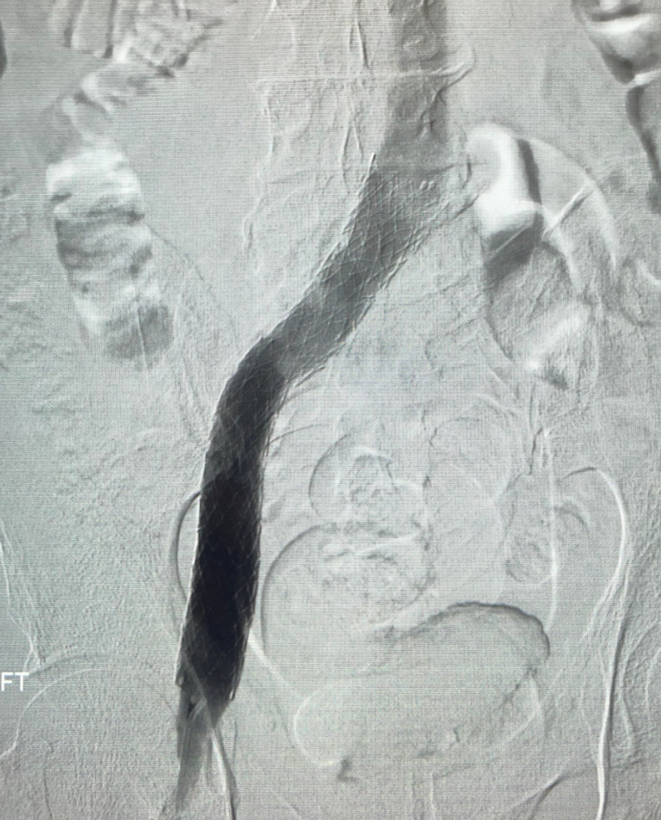
Here’s a pre versus post of our iliac segment after hauling clot:


You can see after clot extraction that we have flow through the iliac segment but there is a clear linear defect proximally and there is contrast reflux into the internal iliac vein. Both findings angiographically suggest evidence of significant May-Thurner.
What next? IVUS. Here’s what I do to make my IVUS as quick and efficient as possible.
- Fix the table in the area of interest and lock it.
- When initiating your IVUS pullback from central to peripheral, have your technologist or nurse who is running the IVUS write down your bookmarks and corresponding locations (my bookmarks are typically IVC, common iliac compression, common iliac reference, external iliac and common femoral).
- Figure out your proximal and distal landing zones while live on the IVUS. Then mark the screen using a dry-erase marker (yes you can overlay, but you want to be precise with this stent and I find it easier for my eyes to look at the stent with a native image). During this time while you are checking out your stent length, have your nurse/tech measure vessel measurements. Please note that it would be rare to use anything other than a 120 or 150 mm length stent.
This entire process of pullback IVUS to stent marking should take less than 10 minutes.
Which stent should you use? Being a millennial IR, I am honestly only really familiar with newer generation venous stents. I have deployed an occasional Wallstent, but they are like Chinese finger traps. If you have the ability to place a newer stent for more precise deployment, take it.
I was the first person in North Carolina to deploy a Vici stent. No one cared and I wasn’t paid a dime for that distinction. Vici’s have since been recalled (sounds like some true pipelayers were undersizing them and they flew to the heart). I then moved over to Venovo by Bard, which was also recalled (faulty deployment mechanism). Now, there are only a couple real options:
- Cook Zilver Vena
- Medtronic Abre
Both are fine stents and work well. I find the Medtronic much easier to deploy as it is a triaxial system (wheel deployment) which for me is easier than the pin-pull mechanism for the Cook Zilver Vena. With either of these stents you size up from the reference common iliac measurement. In our case it measured 14, so I chose a 16. For those in the OBL setting doing chronic DVT work, note that all these stents are pricey, but I think it’s worth the added cost. If you want to know how much, generally a ballpark of $1500 depending on your volume based contract. Note that CMS pays about 3k/venous stenting case in the OBL. Despite popular opinion by hospital based purists, these are not money makers in the OBL even for the most ambitious pipe-layers.
Now this is the part which is just as much of an art as it is science: nailing the iliac confluence. While it is true that you want to use IVUS to identify your confluence, fluoroscpic landmarks can be very helpful. One thing I like to do is to take a Sos and hook it around the confluence. Also understand that for May-Thurner, your true iliac confluence may not be exactly where you want your stent to land. You are trying to oppose the force of the overlying right common iliac artery which will land obliquely to the vessel and may involve the vein somewhat more cranial than you would expect. This is why IVUS is so helpful. From a fluoroscopic standpoint, you want your stent to land between the posterior spinous process and the contralateral pedicle lateral wall (check out this reference). Landing this stent exactly is somewhat of an art and takes a few cases to get down. You will inevitably land one or two a touch short, or a touch long. I’d say err on the side of long so you cover the lesion and take solace in the fact that rates of contralateral DVT are incredibly low for new generation open cell venous stents. Furthermore, keep in mind that back in the day the teaching was to have the Wallstent touch the contralateral IVC.
Here’s our venogram after pre-dilation to 12 mm, and subsequent placement of a 16mm x 150 mm Abre followed by post dilation to 16 mm.
Here’s a CT done a few weeks later (obtained for an unrelated complaint):
I always IVUS after to make sure the stent is well expanded. We close with a purse-string suture about the access. I remove the suture the next morning when rounding.
I place patients on Lovenox for 3 weeks followed by transition to DOAC. After stenting antiplatelet medications are recommended. I go with plavix. I keep patients on Plavix and DOAC for at least 3 months before transitioning to a baby aspirin daily. If you ask 5 different IRs, you’ll likely get 5 different answers regarding anticoagulation and antiplatelet agents. I see patients at 1 month, 3 months, 6 months and annually thereafter with ultrasound at these visits. These are happy clinic visits for the most part. Some of my most grateful patients to this day are those I see in follow-up for in-hospital DVT care.
If you have any thoughts or suggestions, please feel free to share. This is how I approach these cases and has worked well for me now having done dozens of them over the last few years. For the trainees out there, note that I only did 3 of these cases in fellowship. Goes to show that your training does not define you. I am by no means an authority and am always looking to learn new things so please comment and share your thoughts.