You’re probably heard about retrograde pedal access, which is a valuable tool in the fight against critical limb ischemia. But have you heard about antegrade pedal access?
Yes, you’ve heard that right. I’m talking about downsticking a pedal vessel. But why on Earth would you ever want to do that?
Consider the scenario where there is a palpable proximal DP pulse, but a persistent foot wound, with perhaps an incomplete pedal loop. No need to stick the groin when you can achieve a favorable outcome much faster by sticking the ankle. Nothing wrong with sticking to tried and true femoral access. Pedal in my opinion is maximizing our endovascular skillset to get these unique subset of PAD cases done faster and just as safe if not safer than traditional femoral approaches.
Of course before taking any patient to angiography you would have seen a patient in clinic and have conducted proper diagnostic assessments including ultrasound and hopefully associated toe pressures.
I like starting off these cases by gently tying the two feet together on the table to minimize motion. Safely sedate the patient if possible, otherwise fentanyl alone or even local alone in a cooperative patient will suffice.
In order to obtain access, I find it preferable to use a high frequency linear transducer (pediatric probe, or “hockey stick” probe). Scan the foot and look for the dorsalis pedis. Follow it up beyond the ankle into the distal third of the tibia. Be sure to compress the vessel along the way. The moment the artery loses compressibility, that’s when you know you’re too far cranial to safely stick. Arterial access in this location with appropriate intravascular tamponade (such as balloon occlusion from an alternative access point) can result in compartment syndrome.
The ideal spot to stick is right above the tibiotalar joint. Seeing as you’re likely a radiologist you definitely know what that is unlike our vascular surgery and cardiology colleagues (sorry, had to throw a jab there).
Once you pick your spot, go ahead and give some lidocaine around the vessel and give yourself at least 1 cm depth from the skin surface to the anterior wall. For access, I like to use a pedal access kit. The ones by Cook are good, as are the ones by Merit. Take the 21 gauge needle, enter at 30 degrees and carefully get to the anterior surface of the vessel before puncture. Carefully advance your wire into the vessel. If you meet resistance, stop, and re-adjust your needle as necessary. Once you have sufficient wire purchase, gently advance the inner dilator of your micropuncture sheath into the vessel. Give some nitro (200 mcg or so depending on the BP) and do a nice injection in an AP projection (please see the last Technical Tuesday post for tips regarding pedal angiography).
From here you can do your thing. Aggressively heparinize (100 units/kg with target ACT >250). The 4 Fr Merit Prelude Ideal sheaths are my favorite for pedal cases. They are double braided and less expensive than Terumo Glide Slenders. I like to cross using an Asahi Fielder XT wire, IVUS with a 0.014 Phillips and vessel prep with an 0.9 Phillips Laser. I avoid orbital atherectomy below the ankle. You could do it if you have bad calcium, but be careful. The wire bias with 1.25 micro crown can cause vessel wrap and some less than desirable outcomes. Balloon angioplasty with a 0.014 balloon of your choice (I like the CSI Jade balloons). Remember to repeat the IVUS after intervention. Any critical dissections can be treated with the Tack Endovascular scaffold system. Yes, you can use this below the ankle. In fact, I was the first ever to do so (per the device manufacturer. It’s off label and I’m pretty sure no one cares I’m the first, but I have to brag about something and I helped mentor a medical student who won a sweet case competition which was the most meaningful part of it all).
Once you’re done, remove your access and hold gentle manual compression for 5 minutes. Check distal signals and put some nitropaste around the puncture site for good measure. Patient can go home in 45 minutes.
Finally, I should note that I have no conflicts of interest. I believe in the products I use, but that doesn’t mean that there aren’t others out there that can get the job done. The exception being the Tack device which really is unique when it comes to below the ankle scaffolding.
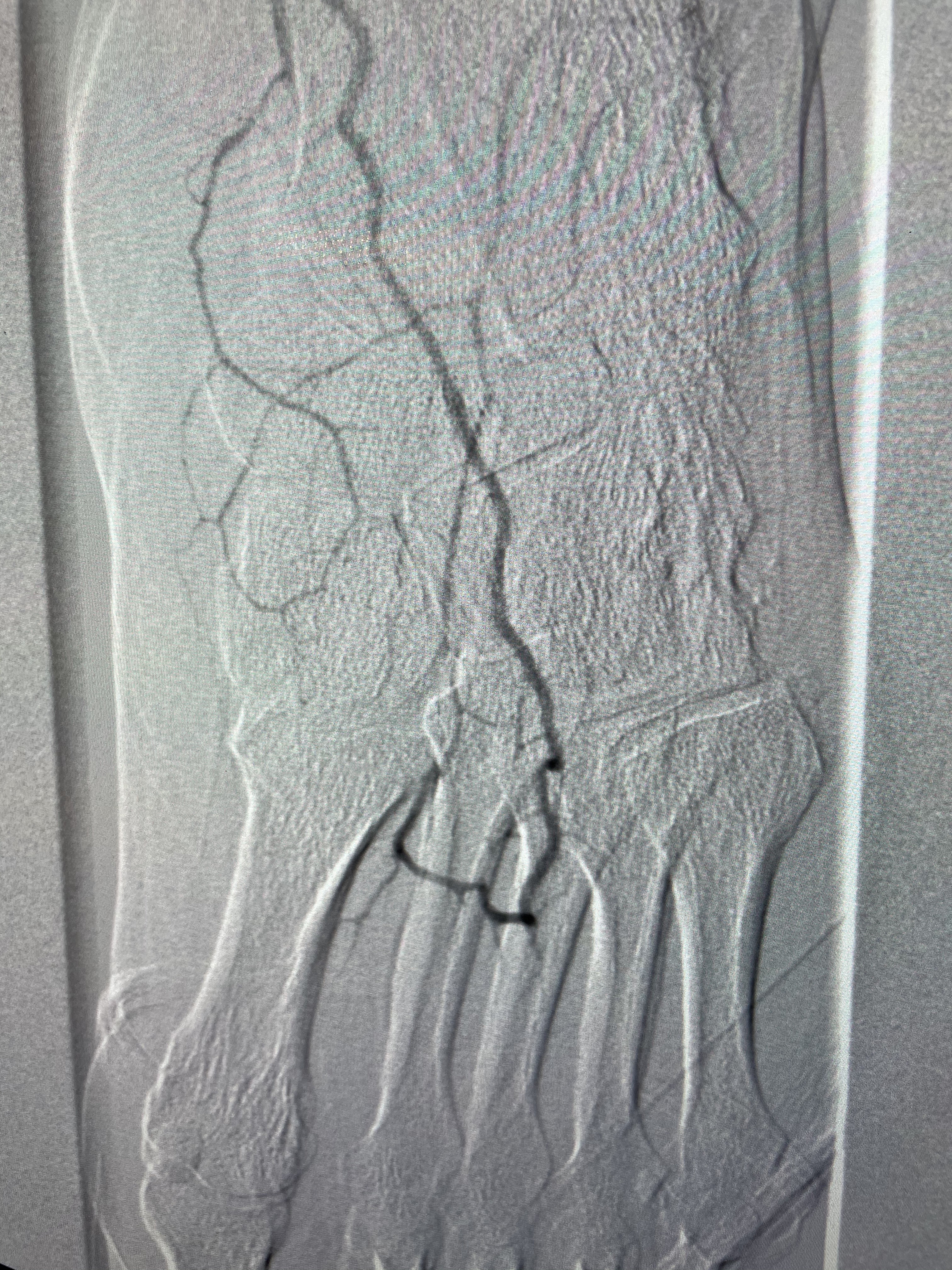
Here’s a nice example of a pre and post distal DP disease with non-healing first digit ulcer, history of COVID-19 (i.e COVID toes):